Non-Invasive Vagus Nerve Stimulation Effects on Brain Waves: A Study of Alpha Power and Stress-Related Attention Signals
Published in: The Society for Neuroscience and The Brain and Behavior journals.
Abstract
Auricular transcutaneous vagus nerve stimulation (tVNS) is a non-invasive technique that delivers mild electrical impulses to a branch of the vagus nerve located in the outer ear. The vagus nerve is a key communication pathway between the body and the brain, influencing mood, and emotions.
In this study, we investigated the acute effects of a single one hour tVNS session in healthy young adults using electroencephalography (EEG), a method that records brainwave activity.
The analysis revealed significant increases in the alpha frequency band (8–12 Hz). Alpha power is typically linked to relaxation and self-related awareness. The increase was observed in regions associated with self-related cognition and tactile sensation. These findings suggest that tVNS can modulate brain oscillatory activity in circuits tied to stress regulation and emotional balance.
Background
The vagus nerve helps regulate stress by shaping attention, mood, and self-related cognition. One major target of vagal activity is a set of midline brain regions engaged during self-reflection and emotion regulation, known as the default mode network (DMN). Because this network is often disrupted when anxiety arises, it serves as an important marker of stress-related brain activity.
In response to stimuli, the brain can generate rapid electrical signals called event-related potentials (ERPs). These can be recorded with an electroencephalograph using an electrode cap (EEG). Two well-studied ERPs are the error-related negativity (ERN), which reflects neural reactions to mistakes, and the visual N2, which reflects attention to visual stimuli. Both signals are often elevated in individuals with heightened stress, and they indicate the brain’s capacity to regulate emotion and maintain cognitive control. These measures can provide sensitive insights into how vagus nerve stimulation can modulate emotional balance at the neural level.
⦁ Design: 1 single tVNS stimulation session of 1 hour using a Roga Device.
⦁ Each participant came to the laboratory once. The experiment contained two phases: Pre-Intervention and Post-Intervention. ⦁ Equipment: - EEG recorded with a 64-channel cap, electrodes placed per int. 10–20 System - Eye blink and movement detection via VEOG and HEOG, converted to bipolar channels offline - Impedance maintained under 5 kΩ - Data collected with 60 Hz notch filter, digitized at 500 Hz
⦁ EEG Processing - Data downsampled to 250 Hz and re-referenced to average in EEGLAB - DC offsets removed, high-pass filtered (1 Hz for spectral analysis, 0.1 Hz for ERP) - Independent component analysis used to remove eye blinks and movements - Ocular and EMG activity flagged and visually confirmed before correction
⦁ EEG Data: - Exported into LORETA for source analysis - Current source densities computed for delta, theta, alpha, beta1, beta2, and gamma frequency bands - Pre and post rest baselines compared with paired t-tests
⦁ ERP Analysis: - Data segmented into stimulus locked and response locked epochs - Epochs baseline corrected and low pass filtered at 30 Hz - N2: measured at FCz, peak amplitude and latency 200–350 ms from incongruent minus congruent trials - ERN: measured at FCz, peak amplitude and latency 0–100 ms from incorrect minus correct trials - Weighted averages based on trial type used for participant waveforms
Results
Alpha Power Reflecting oscillatory activity in the 7.5–14 Hz range, is commonly linked to internal attention, memory, and regulation of stress responses. This range of brain activity is often referred to as the alpha state, a condition associated with relaxed but alert mental processing and reduced external distraction.
In this study, tVNS stimulation led to clear increases in alpha activity within specific areas of the left hemisphere. These effects suggest that the stimulation influenced brain networks tied to stress and attention, supporting the idea that tVNS helped participants shift into an alpha state.
There was a significant increase in alpha power in the left precuneus (BA7) [t=3.20, p<.05] (Figure H1).
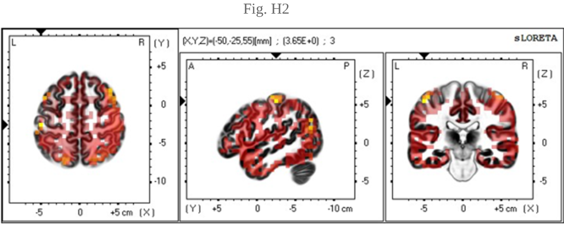
There was also a significant increase in alpha power in the left postcentral gyrus, specifically in the head region (BA1) [t=3.65, p<.05] (Figure H2).
The observed increases were specific to the precuneus and postcentral gyrus. These increases in alpha activity suggest that the tVNS stimulation helped the brain shift toward a calmer, more focused state.
Event Related Potentials (ERPs)
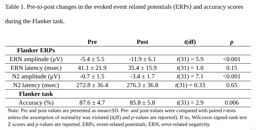
ERPs showed significant increases in ERN and N2 amplitudes, with no changes in their timing. These effects suggest stronger engagement of attention related brain networks.
There were significant pre-to-post increases in the ERN amplitude [t(31)=5.9, p<.001] (Fig. A).
There was also a significant increase in the N2 amplitude [t(31)=7.1, p<.001] (Fig. B).
There was no significant changes on ERN latency [t(31)=1.0, p=.15] or N2 latency [t(31)=.33, p=.65] (Table 1).
There was a significant pre-to-post decrease of ~2% in the correct number of Flanker trials [t(31)=2.9, p=.006] (Table 1).
There were no correlations between the percent of correct responses on the Flanker task and the ERN amplitudes or N2 amplitudes. These changes suggest enhanced activity in the anterior cingulate cortex, improved focus and emotional control.
Theta and High Beta Activity
Theta power rose modestly, consistent with a more relaxed state often observed during meditation, while high beta power increased across both frontocentral and posterior regions, suggesting downregulation of DMN activity.
Theta FrontCentral (4–7 Hz) increased slightly from 5.75 to 6.16, by 7 (p = 0.04) (Fig. C).
High Beta FrontCentral (18–30 Hz) increased from 0.85 to 0.94, a 11% rise (p = 0.02) (Fig. D).
High Beta Posterior (18-30 Hz) increased from 0.03 to 0.04, a 33% rise (p = 0.01) (Fig. E). Increased theta and beta power with reduced activity in the DMN indicates an enhanced state of relaxation and a shift away from stress related rumination.
Conclusion
Our ROI analysis showed increased alpha power in the left parietal lobe, specifically in the precuneus and the head region of the postcentral gyrus. This may indicate an altered activation pattern of the default mode network during the final resting state, reflecting increased capacity for working memory or engagement in autobiographical memories involving other people.
Increased alpha power in the head region of the left primary somatosensory cortex may reflect a change in tactile perception resulting from electrode placement and stimulation. The left hemisphere has greater sensitivity than the right in this region, suggesting the same may apply to the head region.
Such increases in alpha activity are consistent with a shift into an alpha state, a condition marked by relaxed internal attention and reduced external distraction, which aligns with the observed modulation of stress and attention networks.
The human brain remains highly complex, and these findings represent only an early step in mapping how vagus nerve stimulation influences its networks. Further studies with larger cohorts, extended protocols, and multimodal measures will be essential to deepen understanding and validate the therapeutic potential of these effects.

.png)
.png)
.png)
.png)
.png)


